




















Most people learn about heart valve prolapse the same way: a stethoscope catches a faint click during a routine physical, a follow-up echocardiogram lands on the schedule, and suddenly the word “prolapse” is attached to their heart. The reaction is usually some mix of alarm and confusion.
The reassuring part is that heart valve prolapse, especially when it involves the mitral or tricuspid valve, is often manageable and frequently causes no symptoms at all. The part worth taking seriously is that a smaller subset of cases progresses, and knowing which features matter helps separate routine monitoring from the rare situations that need a closer look.
The condition has a long and somewhat tangled history in American cardiology. For decades, mitral valve prolapse was overdiagnosed by stethoscope alone, then redefined with more precise echocardiographic criteria.
The result today is a clearer picture: prolapse is common, usually mild, and rarely the cause of serious cardiac events. What follows is a practical guide to what heart valve prolapse actually is, how it shows up, what testing looks like, and how cardiologists decide between watchful waiting and active treatment.
- Heart valve prolapse means one or both valve flaps bulge backward when the heart contracts, most often affecting the mitral valve and less commonly the tricuspid.
- Many people have no symptoms; when they appear, palpitations, chest discomfort, fatigue, and shortness of breath are the most common.
- Most cases need only periodic monitoring, but worsening regurgitation, persistent symptoms, or arrhythmias can warrant medication or valve repair.
Read More: Exercise for Heart Failure Patients: What’s Safe and Effective
What Is Heart Valve Prolapse?
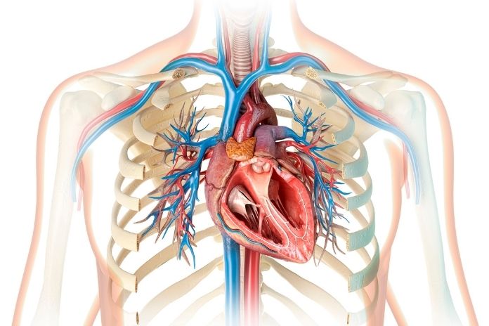
Blood moves through the heart in a strict one-way pattern, and the valves are what keep it that way. The mitral valve sits between the left atrium and left ventricle, and the tricuspid sits between the right atrium and right ventricle. Each one opens to let blood through and snaps shut to prevent it from sloshing backward when the ventricle squeezes.
The leaflets themselves are anchored by thin fibrous cords called chordae tendineae, which tether the valve to small muscles in the ventricular wall and keep the seal tight under pressure.
In prolapse, the valve leaflets are too floppy or too stretched, so when the ventricle contracts, they bow upward into the atrium instead of sealing flat. Mild prolapse may not let any blood through in the wrong direction. More significant prolapse creates an imperfect seal, and blood leaks backward, a problem called mitral valve regurgitation when it occurs on the left side.
According to a landmark Framingham Heart Study analysis published in the New England Journal of Medicine, classic mitral valve prolapse affects roughly 2.4 percent of adults in the general population. Mitral valve prolapse is far more common, partly because the left side of the heart works against higher pressures and the mitral leaflets carry more mechanical load over a lifetime.
Tricuspid valve prolapse exists but is rare on its own; tricuspid valve disease more often shows up as functional regurgitation tied to right-sided heart pressures or a stretched tricuspid annulus, rather than primary leaflet prolapse.
The diagnostic workup overlaps, but the right-sided anatomy and the company of the tricuspid valve keep with the lungs and venous return, give it a different clinical flavor. Symptoms also tend to differ.
Mitral disease leans toward palpitations and exertional breathlessness, while significant tricuspid regurgitation more often shows up as swelling in the legs, abdominal fullness, and visible neck vein pulsations, signs that blood is backing up into the body’s venous system rather than into the lungs.
Common Symptoms of Mitral and Tricuspid Valve Prolapse
The symptoms list is short and familiar to anyone who has dealt with a vague cardiac complaint:
- Palpitations, often described as fluttering, skipping, or a brief hard thump
- Chest discomfort that does not behave like classic angina
- Shortness of breath, particularly with exertion
- Fatigue or a noticeable drop in exercise tolerance
- Dizziness or lightheadedness, sometimes when standing quickly
Mild prolapse, with no meaningful regurgitation, often produces nothing the patient can feel. Blood still moves forward efficiently, the chambers do not enlarge, and the rhythm stays steady. Plenty of people walk into a cardiology office for unrelated reasons and find out they have prolapse only because an imaging study picked it up.
When regurgitation worsens, the picture changes. Swelling in the legs or abdomen, shortness of breath at rest or while lying flat, and a clearly irregular heartbeat are signs that the valve is failing to hold blood back. These are not symptoms to wait out. They suggest the heart is working harder than it should and that imaging should be repeated sooner rather than later.
Read More: Supplements That Can Cause Heart Palpitations: What You Should Know to Stay Safe
What Causes Heart Valve Prolapse?

Valve tissue is not static. Over time, the leaflets can undergo myxomatous degeneration, a process in which the connective tissue weakens and the leaflets thicken and stretch. This is the most common pathology behind primary mitral valve prolapse and explains why the condition often becomes more apparent in middle age.
A genetic component runs through many cases. The condition can cluster in families, and it shows up much more often in people with inherited connective tissue disorders such as Marfan syndrome and Ehlers-Danlos syndrome.
Dr. Robert A. Levine, MD, a senior physician in the Cardiac Ultrasound Laboratory at Massachusetts General Hospital, has spent decades studying the genetic underpinnings of the disease. As he put it in a Mass General research video, his team is focused on “discovering the genes causing mitral valve prolapse, the most common cause for operation for mitral regurgitation.”
Structural heart changes, including dilation of the left ventricle or the valve annulus, can pull on the leaflets and exaggerate prolapse. Rarely, prolapse develops or worsens after infective endocarditis, blunt chest trauma, or rheumatic heart disease. A comprehensive review in Circulation by Delling and Vasan summarized how genetics, leaflet biology, and ventricular geometry interact across the spectrum of MVP severity.
How Doctors Diagnose Valve Prolapse
The classic auscultatory finding for mitral valve prolapse is a mid-to-late systolic click, sometimes followed by a murmur when regurgitation is present. The click is the sound of the valve leaflets snapping into their billowed position. A skilled clinician can pick it up with a basic stethoscope, though the finding is more subtle than people assume.
Echocardiography is the workhorse for confirming valve prolapse. A transthoracic echocardiogram uses sound waves to image the leaflets in motion, measure how far they bow into the atrium, and quantify any backward blood flow.
When more detail is needed, a transesophageal echocardiogram gives a sharper view by placing the transducer in the esophagus directly behind the heart. An electrocardiogram is often done to check for arrhythmias. A Holter monitor, worn for 24 to 48 hours or longer, can capture palpitations that come and go.
Cardiac MRI is reserved for select cases, including patients with suspected myocardial scarring or a need to clarify the severity of regurgitation. The decision about which tests to add depends on symptoms, exam findings, and how cleanly the echo answers the clinical question.
Read More: Heart Murmur vs. Irregular Heartbeat: What’s the Difference?
When Is Mitral or Tricuspid Valve Prolapse Serious?
The severity of prolapse matters less than the severity of valve leakage. A leaflet that bows a few millimeters too far but seals adequately is a different problem from one that allows a steady stream of blood backward into the atrium. Cardiologists grade regurgitation as mild, moderate, or severe, and that grading drives almost every decision that follows.
Worsening regurgitation can enlarge the left atrium and left ventricle over time, raising the risk of atrial fibrillation and, in advanced cases, heart failure. A rare subset of patients with mitral valve prolapse experience complex ventricular arrhythmias and an elevated risk of sudden cardiac death.
A pivotal study by Basso and colleagues in Circulation examined the pathology of young adults who died suddenly with MVP and found a consistent pattern of myocardial fibrosis at the papillary muscles and inferobasal wall, pointing to a structural substrate for these arrhythmias. Patients with moderate to severe regurgitation, frequent ventricular ectopy, bileaflet prolapse, or mitral annular disjunction generally warrant more frequent follow-up.
Dr. Francesca Nesta Delling, MD, MPH, medical director of the Echocardiography Laboratory at UCSF, has been outspoken about not dismissing every case as harmless. “Mitral valve prolapse should not be underestimated as being mostly benign,” she said in a UCSF Cardiology profile, noting that better tools are needed to flag the small group at higher risk.
Treatment Options for Heart Valve Prolapse
For most patients, treatment is observation. A routine echocardiogram every few years is enough if there is no significant regurgitation. People with mild regurgitation are typically followed annually. Limiting stimulants such as excess caffeine and nicotine can reduce palpitations, and staying well hydrated helps blunt the dizziness some patients describe.
When symptoms are bothersome, beta blockers are the first medication most cardiologists try. They slow the heart rate, soften the force of contractions, and often quiet palpitations. Diuretics are added if fluid retention develops, and anticoagulants enter the picture if atrial fibrillation appears.
It is important to be clear that none of these medications fixes the prolapse itself; they manage symptoms and complications. Surgery enters the conversation when regurgitation becomes severe, when symptoms persist despite medical therapy, or when the heart chambers start to enlarge. The preferred approach today is valve repair rather than replacement whenever the anatomy allows it.
Dr. A. Marc Gillinov, MD, Chairman of the Department of Thoracic and Cardiovascular Surgery at Cleveland Clinic, is one of the field’s most prominent voices on this point. “When repair is feasible, it is the best option for degenerative mitral valve disease,” he said in a Cleveland Clinic Consult QD article, noting that repair restores normal life expectancy in ways replacement often does not.
Treatment Approach
Typical Indication & Goals
| Treatment Approach | Typical Indication | Goal |
|---|---|---|
| 🩺 Routine Monitoring | Mild prolapse with no or trace regurgitation | Detect progression early |
| 💊 Beta Blockers | Palpitations, anxiety, or mild arrhythmias | Symptom relief |
| 💧 Diuretics | Fluid retention or mild heart failure symptoms | Reduce volume overload |
| 🩸 Anticoagulants | Atrial fibrillation | Stroke prevention |
| 🫀 Valve Repair Surgery | Severe regurgitation, symptoms, or chamber enlargement | Restore valve function |
| 🔄 Valve Replacement | Repair not feasible or valve severely damaged | Replace failed valve |
The table above is a simplified map, not a prescription. The right treatment for any individual depends on echocardiographic findings, symptoms, and overall cardiac function, and the decision is made jointly between the patient and cardiologist.
Living With a Prolapsed Heart Valve

Most people with mitral or tricuspid valve prolapse can stay fully active. Aerobic exercise, strength work, and recreational sports are usually fine, and avoiding activity is more often a mistake than a precaution.
Competitive athletes and people with severe regurgitation are different cases and need individualized clearance, particularly if there is any history of fainting, complex arrhythmias, or significant chamber enlargement.
Pregnancy is generally well tolerated in people with mild mitral or tricuspid valve prolapse. The added blood volume and cardiovascular demand can occasionally trigger palpitations or make a faint murmur louder, but most pregnancies proceed without complication.
Women with moderate to severe regurgitation should be evaluated by a cardiologist before pregnancy when possible, since closer monitoring is helpful, and a small number of cases require treatment adjustments.
The long-term outlook is reassuring for the majority. Mild prolapse remains stable for decades in many people, and life expectancy is essentially unchanged. A subset progresses, which is why follow-up matters even when nothing feels wrong.
A community-based study published in JACC Cardiovascular Imaging by Topilsky and colleagues found that moderate or greater tricuspid regurgitation, while sometimes silent, carried meaningful long-term mortality risk, a reminder that even right-sided valve disease deserves serious attention once it crosses a certain threshold.
Patients tend to do better when they engage with their condition rather than ignore it, and that engagement is usually as simple as showing up for the scheduled echo.
When to See a Doctor About Possible Valve Prolapse
Some symptoms warrant a same-day or urgent visit. Fainting, severe shortness of breath, persistent chest pain, and a rapid or irregular heartbeat that lasts more than a few minutes are all signals to be evaluated promptly, regardless of a known prolapse diagnosis.
New or worsening leg swelling, especially with shortness of breath when lying down, can point to advancing regurgitation.
Questions to Ask During a Cardiology Visit
A productive cardiology visit is built on specific questions. Ask about the severity of the prolapse on imaging, whether any regurgitation is present and how severe it is, how often imaging should be repeated, and whether any activity adjustments are needed. For people with tricuspid involvement, ask whether right-heart pressures or right ventricular function is normal.
Dr. Patrick T. O’Gara, MD, Director of Strategic Planning for the Cardiovascular Division at Brigham and Women’s Hospital, led the writing of the 2025 ACC expert consensus on tricuspid regurgitation.
The document was designed, in his words, “to enable the clinician to assimilate the most important developments in this dynamically evolving field to improve the care of patients with severe TR,” a useful reminder that tricuspid disease has moved out of the shadow of mitral valve management.
Read More: 8 Heart Warning Signs During Exercise You Should Never Ignore, According to Cardiologists
Key Takeaway on Mitral and Tricuspid Valve Prolapse
Heart valve prolapse is one of the most common findings on a cardiac echo, and for most people, it remains a footnote in their medical record rather than a defining diagnosis. Mitral valve prolapse is far more frequent than tricuspid involvement, and both conditions tend to behave well when regurgitation is mild and the heart chambers stay normal in size.
The medical literature has shifted in recent decades. Once viewed as universally benign, mitral valve prolapse is now understood as a spectrum, and a small but real subset of patients carries a higher risk for arrhythmias or progressive regurgitation. That is not a reason for panic.
It is a reason for periodic checkups, attention to new symptoms, and a relationship with a cardiologist who knows the patient’s baseline. The practical message for anyone who has just been told they have heart valve prolapse is straightforward. Keep your appointments, report new symptoms, stay active, and ask specific questions about your imaging results.
Most people with mitral or tricuspid valve prolapse live full, unrestricted lives, and the small minority who need more do far better when their disease is caught early.
References
- Basso, C., Perazzolo Marra, M., Rizzo, S., De Lazzari, M., Giorgi, B., Cipriani, A., Frigo, A. C., Rigato, I., Migliore, F., Pilichou, K., Bertaglia, E., Cacciavillani, L., Bauce, B., Corrado, D., Thiene, G., & Iliceto, S. (2015). Arrhythmic mitral valve prolapse and sudden cardiac death. Circulation, 132(7), 556–566.
- Cleveland Clinic. (2024). Mitral valve prolapse.
- Delling, F. N., & Vasan, R. S. (2014). Epidemiology and pathophysiology of mitral valve prolapse: New insights into disease progression, genetics, and molecular basis. Circulation, 129(21), 2158–2170.
- Freed, L. A., Levy, D., Levine, R. A., Larson, M. G., Evans, J. C., Fuller, D. L., Lehman, B., & Benjamin, E. J. (1999). Prevalence and clinical outcome of mitral-valve prolapse. New England Journal of Medicine, 341(1), 1–7.
- Johns Hopkins Medicine. (2024). Mitral valve prolapse.
- Mayo Clinic. (2024). Mitral valve prolapse: Diagnosis and treatment.
- Topilsky, Y., Maltais, S., Medina Inojosa, J., Oguz, D., Michelena, H., Maalouf, J., Mahoney, D. W., & Enriquez-Sarano, M. (2019). Burden of tricuspid regurgitation in patients diagnosed in the community setting. JACC: Cardiovascular Imaging, 12(3), 433–442.
- Mayo Clinic. (n.d.). Mitral valve prolapse – Symptoms and causes.
- Cleveland Clinic. (n.d.). Mitral valve prolapse.
- Johns Hopkins Medicine. (n.d.). Mitral valve prolapse.
- Medtronic. (n.d.). Mitral valve disease.
- Mass General Brigham. (n.d.). Mitral valve prolapse.
- Guy’s and St Thomas’ Specialist Care. (n.d.). Mitral valve problems: Signs, diagnosis and treatment.
- UCSF Department of Surgery. (n.d.). Mitral valve prolapse.
In this Article