

















When your doctor mentions ordering a cardiac stress test, it helps to know what that actually means, and whether a standard stress test or a nuclear stress test is the right call for your situation.
Both evaluate how your heart handles physical demand, but they work differently and answer different questions. A standard stress test tracks your heart’s electrical activity during exercise.
A nuclear stress test adds cardiac imaging with a radioactive tracer to show how well blood flows to the heart muscle. Understanding the difference helps you walk into the conversation with your doctor prepared.
- Standard stress tests check heart rhythm during exercise
- Nuclear tests show blood flow using imaging.
- Doctors typically start with basic testing and move to nuclear scans for a clearer picture or when standard results are unclear.
- Your symptoms, risk factors, and ability to exercise all factor into which test fits your needs.
Read More: What Not to Do Before a Stress Test: Doctor-Approved Preparation Guide
What Is a Cardiac Stress Test?
A cardiac stress test shows how the heart performs under physical demand. Because the heart pumps harder and faster during exercise, the test can reveal changes in blood flow that aren’t visible at rest.
Doctors use the results to check for coronary artery disease, evaluate unexplained symptoms like chest pain or shortness of breath, and monitor patients with known heart conditions.
How a Standard Stress Test Works
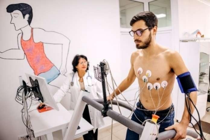
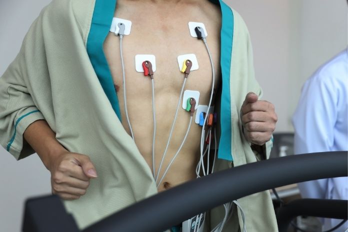
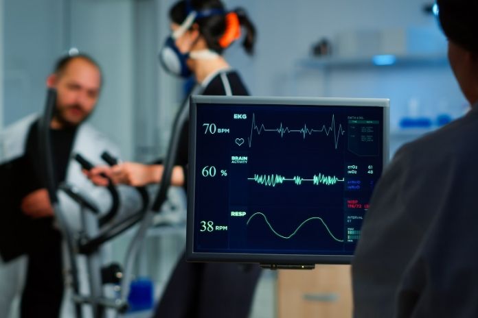
For a standard exercise stress test, you walk on a treadmill or pedal a stationary bike while a technician gradually increases the speed or resistance. Before you start, the care team attaches ECG electrodes to your chest to track your heart’s electrical activity throughout the test. They also monitor your blood pressure and heart rate at each stage.
The test typically takes 30 to 60 minutes from start to finish, including prep and recovery. You’ll need to avoid caffeine and certain medications beforehand. Your doctor will give you specific instructions. If you can’t exercise sufficiently, medication can be given through an IV to mimic the effects of physical exertion on the heart.
What a Nuclear Stress Test Adds
A nuclear stress test, also called a myocardial perfusion scan, layers cardiac imaging on top of the standard exercise protocol. A radioactive tracer is injected through an IV, travels through your bloodstream, and is absorbed by the heart muscle. A gamma camera then captures images showing where blood is flowing normally and where it may be reduced or blocked.
Doctors take two sets of images: one while your heart is at rest and one after it has been stressed. Comparing those images can reveal areas of reduced blood flow, signs of prior heart damage or scarring, and information about your heart’s pumping function. The whole process typically takes two to four hours.
Stress Test vs. Nuclear Stress Test: Key Differences
Both tests evaluate heart health, but they measure different things and suit different clinical situations.
Cardiac Testing Guide
Standard Stress Test vs Nuclear Stress Test
| Category | Standard Stress Test | Nuclear Stress Test |
|---|---|---|
| What it measures | Heart rhythm, electrical activity, exercise capacity | Blood flow to heart muscle, perfusion defects |
| Imaging | No | Yes (radioactive tracer + gamma camera) |
| Duration | 30–60 minutes | 2–4 hours |
| Prep | Avoid caffeine; some meds withheld | Fast for several hours; avoid caffeine; some meds withheld |
| Radiation | None | Yes (approx. 8–11 mSv) |
| Best for | Initial screening in lower-risk patients | Higher-risk patients, unclear ECG results, post-treatment monitoring |
Accuracy for Detecting Blocked Arteries
For detecting coronary artery disease (CAD), the two tests are not equally powerful. A large meta-analysis found the exercise ECG has a pooled sensitivity of about 68% and specificity of 77% for identifying CAD.
Nuclear imaging improves on that, with studies reporting sensitivity around 83% and specificity around 77%. Put simply, the nuclear test catches more cases of reduced blood flow that the standard ECG might miss, but neither test is definitive on its own. Results always need to be read alongside your symptoms and full medical history.
Radiation Exposure Explained
Because the nuclear test uses a radioactive tracer, patients are exposed to ionizing radiation — typically between 8 and 11 millisieverts (mSv) for a standard technetium-based scan. For comparison, the average American receives about 3 mSv per year from natural background sources, and a standard chest X-ray delivers roughly 0.02 mSv.
The radiation clears your body within a few days, and drinking plenty of water speeds that process. For most patients, the diagnostic benefit far outweighs the small radiation risk. Pregnant women should not have nuclear imaging, and patients undergoing frequent radiation-based procedures should discuss cumulative exposure with their doctor.
Read More: Coronary Artery Disease – Causes, Symptoms, Diagnoses And Treatment
Who May Need a Standard Stress Test?
Doctors often start with an exercise stress test for:
- Symptom evaluation: Chest pain, shortness of breath, palpitations, dizziness, or unexplained fatigue during activity.
- Cardiovascular risk screening: Patients with high blood pressure, high cholesterol, diabetes, smoking history, or a strong family history of heart disease.
- Known cardiac conditions:People with coronary artery disease or arrhythmias who need routine monitoring.
- Pre-surgical clearance: Before major surgery, a stress test can help gauge whether the heart can handle the added physical load.
- Exercise clearance: Adults over 40 with risk factors who are starting a vigorous exercise program.
The standard test works best when a patient can exercise adequately and has a readable baseline ECG. For lower-to-intermediate risk patients who check both boxes, it’s often the right first step.
Who May Need a Nuclear Stress Test?
A nuclear cardiac stress test is typically recommended when a standard stress test isn’t sufficient or isn’t possible. Warning signs of a more serious cardiac problem, chest pain, shortness of breath, fatigue, or dizziness, may point your doctor toward nuclear imaging for a more complete picture.
Specific situations where a nuclear test is preferred include:
- Higher-risk patients: Those with a prior heart attack, diabetes, multiple cardiovascular risk factors, or known coronary artery disease (CAD).
- Abnormal baseline ECG: Conditions like left bundle branch block, a pacemaker rhythm, or certain medication effects can make standard ECG readings unreliable. Nuclear imaging bypasses that limitation.
- Inconclusive standard test results: If an exercise stress test doesn’t give a clear answer, nuclear imaging can identify whether reduced blood flow is actually causing the problem.
- Post-treatment monitoring: After a stent, bypass surgery, or other cardiac procedure, a nuclear scan helps doctors assess whether the treatment worked and whether blood flow has improved.
Patients Who Can’t Exercise Normally
Some patients can’t reach an adequate heart rate on a treadmill due to arthritis, mobility issues, lung disease, or other conditions. In those cases, doctors use a pharmacologic stress test: a medication given through an IV that mimics exercise by stressing the heart.
The most common agents are vasodilators like adenosine or regadenoson, which increase blood flow through the coronary arteries and reveal differences between healthy and narrowed vessels. Dobutamine is used when vasodilators aren’t appropriate, such as in patients with severe lung disease. The imaging process is the same regardless of how the heart is stressed.
Read More: Heart Murmur vs. Irregular Heartbeat: What’s the Difference?
What Happens During a Nuclear Stress Test?
- IV placement and resting images: A technician places an IV and injects the radioactive tracer. After it circulates through your system (usually 15–30 minutes), the gamma camera takes the first set of images while your heart is at rest.
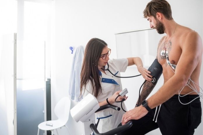
- Exercise or pharmacologic stress: ECG electrodes go on your chest and abdomen so the team can monitor your heart rate throughout. If you’re physically able, you’ll walk on a treadmill with the speed and incline increasing gradually. If not, the stress medication is delivered through the IV.
- Stress images: Once your heart reaches the target level of exertion, the second round of imaging begins. These images capture how your heart responds under stress.
- Recovery monitoring: You’ll stay connected to the ECG for a period after the test so the team can watch your heart rate and rhythm return to baseline.
What the Images Can Reveal
Doctors compare the resting and stress images side by side. An area that shows reduced blood flow only during stress suggests a blockage that limits supply when the heart works hard, a sign of coronary artery disease.
An area with reduced flow at both rest and stress may indicate prior heart damage or scar tissue from a past heart attack. The scans can also show information about your heart’s ejection fraction, a measure of how efficiently it pumps.
Understanding Stress Test Results
After the test, your doctor interprets the results in one of four ways:
- Normal (negative): Your heart showed adequate blood flow and a healthy rhythm throughout activity. This generally means significant coronary blockage is unlikely, though your doctor will weigh results against your full clinical picture.
- Abnormal (positive): The test detected changes, such as ECG irregularities, a drop in blood pressure, chest discomfort, or reduced blood flow on imaging, suggesting the heart wasn’t receiving enough oxygen during exertion. Additional testing or treatment may follow.
- Inconclusive: The test showed changes that don’t clearly point to ischemia. Something may be happening, but more information is needed before drawing conclusions.
- Uninterpretable: The test couldn’t be completed, for example, due to equipment issues or symptoms that forced an early stop. An alternative stress test approach may be scheduled.
Why False Positives and False Negatives Happen
No stress test is perfect. The standard exercise ECG misses some cases of CAD (false negatives) and can flag problems that don’t exist (false positives), particularly in women of childbearing age, where false positive rates are higher.
Nuclear imaging is more accurate, but it still carries some margin of error. Results are most reliable when interpreted alongside your symptoms, risk profile, and, if needed, follow-up imaging such as a stress echocardiogram or coronary angiography.
Risks and Side Effects of Stress Testing
Stress tests are generally safe and performed under continuous medical supervision with emergency equipment nearby. If complications do arise, they can include:
- Lightheadedness or dizziness
- Nausea
- Low blood pressure
- Shortness of breath or wheezing (particularly with pharmacologic agents)
- A fast or irregular heartbeat, which usually resolves once exertion stops
- Skin irritation around ECG electrode sites
- For nuclear tests: a mild allergic reaction to the radioactive tracer (uncommon)
- Chest discomfort or pressure
- Heart attack (very rare)
The care team monitors you throughout the test and during recovery to respond quickly to any of these.
Read More: How to Detect Silent Heart Attacks and Reduce Your Risk
How Doctors Decide Which Test You Need
Factors That Influence Test Selection
Physicians weigh several factors when choosing between a stress test vs. nuclear stress test:
- Symptoms: The nature, frequency, and severity of chest pain, shortness of breath, or other cardiac symptoms.
- Age and cardiovascular risk: Older patients and those with multiple risk factors are more likely to need nuclear imaging.
- Ability to exercise: Patients who can’t exercise adequately will need a pharmacologic protocol, typically paired with nuclear imaging.
- Baseline ECG: An abnormal resting ECG often makes nuclear or echocardiographic imaging necessary.
- Prior cardiac history: A previous heart attack, stent, or bypass surgery typically calls for nuclear imaging to assess current perfusion.
A Note on Cost
Cost is a real factor for many patients. A standard treadmill stress test generally runs $200–$500 without insurance.
A nuclear stress test is considerably more expensive, often $1,500–$4,000 or more depending on the facility and location, because it involves the radioactive tracer, specialized imaging equipment, and longer appointment time.
Most major insurers cover both tests when medically indicated. Medicare Part B covers nuclear stress tests when ordered as medically necessary and typically pays 80% of the approved amount after your Part B deductible.
If cost is a concern, ask your doctor whether a standard stress test is an appropriate starting point. In many cases, it is.
Questions to Ask Before Scheduling a Test
- What is this test looking for specifically?
- Does my situation require cardiac imaging, or will ECG monitoring be enough?
- How should I prepare? Any medications to avoid, fasting requirements?
- What follow-up might I need based on the results?
Conclusion
The stress test vs. nuclear stress test comparison ultimately comes down to what your doctor needs to know and what your health situation requires. A standard exercise stress test is a solid, affordable first step for evaluating heart rhythm, exercise tolerance, and initial coronary artery disease risk in lower-to-intermediate risk patients.
A nuclear stress test delivers more detailed information, specifically about blood flow to the heart, and is the better choice when the standard test isn’t sufficient, isn’t possible, or leaves key questions unanswered.
If you’ve been told you need a cardiac stress test, the right conversation to have is with your cardiologist or primary care doctor. Your symptoms, medical history, risk factors, and ability to exercise all shape which test gives the most useful picture of your heart health.
The goal of both is the same: catching problems early and giving you and your doctor the information needed to protect your heart.
Read More: Balance Your Stress: The Top 6 At-Home Cortisol Tests for Stress Management
References
- American Heart Association. What is a stress test?
- Mayo Clinic. (2025, January 7). Stress test
- Oklahoma Heart Hospital. (2022, February 19). What to expect during a cardiac stress test.
- Liv Hospital. Difference between a stress test and a nuclear stress test.
- South Shore Cardiovascular Associates. Nuclear stress test: What you can expect.
- The Heart Clinic. (May 03, 2024). Here’s How to Interpret the Results of Your Exercise Stress Test.
- Stanford Health Care. Risks of stress test.
In this Article