


")
")


The way physicians identify and treat cardiovascular diseases has been revolutionized by advanced cardiac imaging. The cardiac CT scan and cardiac MRI are among the most powerful tools available today. These tests are part of modern heart imaging tests that allow physicians to see detailed pictures of the heart, blood vessels, and surrounding structures without surgery.
This article covers the definition, operation, and applications of these scans. You will discover what a cardiac CT scan shows, what a cardiac MRI is, and how medical professionals choose between a cardiac CT vs. MRI for diagnosis.
- Cardiac CT quickly detects artery blockages and calcium buildup using X-rays.
- Cardiac MRI provides detailed images of heart structure, function, and tissue without radiation.
- Doctors choose between them based on symptoms, heart rate, and diagnostic needs.
Read More: MRI Scans Show Exercise Can Make Your Brain Look Younger
What Are Cardiac CT Scans and Cardiac MRIs?
Common imaging examinations used to assess the heart include cardiac CT and cardiac MRI studies. Each diagnostic test’s findings can assist medical professionals in identifying and diagnosing a range of cardiac conditions. Each test yields detailed images of the heart, but their applications and goals differ.
Every test operates differently and is more appropriate in particular circumstances.
Cardiac CT Scans: CT (Computerized Tomography) – Cardiac CT scans create detailed three-dimensional images of the heart and blood vessels. Patients who are pregnant, have a high risk of cancer, or are undergoing radiation therapy are generally not good candidates for
CT scans involve low-level radiation. For these patients, an MRI usually offers better diagnostic information. Most patients will need a contrast dye injection to help improve their images. An injection of contrast dye is usually safe for most patients; however, some patients may experience a negative reaction to the contrast. Some patients may also require beta blockers before their imaging to help slow their heart rate.
Cardiac MRIs: In a cardiac MRI, a powerful magnet, radio frequencies, and a computer create detailed still and moving images of the heart for diagnostic purposes. This safe, non-invasive technique scans the body to create moving images of the beating heart, as well as data on its structure and its veins.
It enables the medical professionals to identify any anomalies in the heart’s chambers, disturbances in blood flow through the heart, and cardiovascular anomalies, such as cardiac tumors, heart valve disease, cardiac hypertrophy (enlarged heart), and other conditions.
What Does a Cardiac CT Scan Show?
A noninvasive, painless examination called a cardiac computed tomography (CT) scan assesses your heart and surrounding blood vessels to find any blockages or artery narrowing. Additionally, a cardiac CT scan can detect abnormalities in your heart’s pumping action, which may indicate heart disease.
A cardiac CT scan can be completed quickly, typically in less than 10 minutes, unlike stress tests. Cardiac CT scans come in two primary varieties:
- Coronary CT angiograms use contrast (dye) to examine the heart’s arteries and identify blockages that may require medical attention.
- A person’s future cardiac risks can be determined by calcium-scoring CT scans, which detect calcium accumulation in the arteries. This test doesn’t require contrast dye or an intravenous (IV) line, and it uses very little radiation.
What Does a Cardiac MRI Show?

A cardiac MRI can check for several issues, such as:
- Heart disease
- Masses in the heart
- Valve disease
- A cardiac clot of blood
- Acute heart damage, such as myocarditis
Cardiologists can use it to assess arrhythmias (abnormal heart rhythms). They can also use it to develop nonsurgical treatments for arrhythmias, such as ablation.
“Cardiac MRI is considered the gold standard for evaluation of heart size and function,” says Dr. Bradley Allen, MD, radiologist. He adds, “In addition, MRI is excellent at tissue characterization and can provide information about scar or inflammation to the heart muscle.”
Read More: What Is a HIDA Scan? Purpose, Procedure, and What It Diagnoses
Cardiac CT vs. MRI: What’s the Difference?
Patients can better understand why doctors choose one test over another by understanding the differences between cardiac CT and MRI:
Radiation and Safety: To create images, cardiac MRI uses radio waves, strong computers, and magnetic fields. In contrast, cardiac CT creates a complete three-dimensional computer model of the heart and its surrounding organs by acquiring X-rays from multiple angles.
Intravenous Contrast: To define the blood-tissue interface and distinguish between various tissue types, cardiac CT requires an intravenous injection of contrast medium. Although cardiac MRI does not require IV contrast, physicians use it in specific situations to highlight problems that would otherwise go undetected.
Heart Rate Control: Both CT and MRI require a reasonably consistent heart rate to produce blur-free images of the beating heart. With a CT scan, that should ideally be 60 beats per minute. Rapid heart rates are generally not a major problem for cardiac MRI as long as the pulse remains regular, whereas they are harmful for cardiac CT.
Patient-Scanner Compatibility: Before a patient undergoes a Cardiac MRI, healthcare providers thoroughly screen for ferromagnetic materials in or on the body because the procedure uses powerful magnets. Fortunately, if a patient has good renal function and is not highly allergic to iodinated contrast media, they can safely undergo cardiac CT with implanted ferromagnetic devices.
Why a Doctor May Recommend Advanced Heart Imaging

Doctors recommend heart imaging tests for a variety of reasons. Chest discomfort, suspected coronary disease, or abnormal test results are common scenarios. When evaluating arrhythmia, unexplained dyspnea, or monitoring established problems, a cardiac CT scan or cardiac MRI may be utilized.
To evaluate damage after a heart attack, these tests are equally crucial. They play a major role in modern MRI for heart disease diagnosis and early detection strategies.
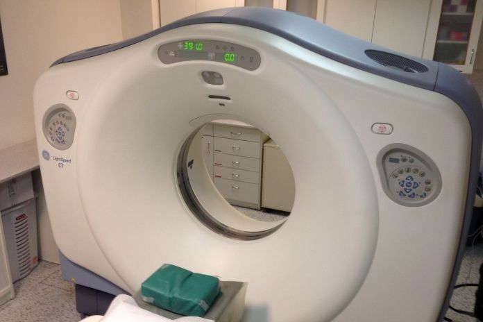
What to Expect During a Cardiac CT Scan
Typically, the complete procedure takes less than an hour. What to anticipate throughout this period is as follows:
- The nurse will insert an intravenous (IV) line into your arm and provide a specific dye (contrast) if you are undergoing a coronary CT angiography. The contrast used in the test will display blood flow in your heart’s arteries and blood vessels, as well as any plaque or calcium accumulation that may reduce blood flow. Because of the contrast, you might taste a metallic note. If you are having a calcium score test, you won’t have an IV or dye.
- A few tiny, sticky electrodes will be applied to your chest by a member of your care team.
- A table will move inside a circular CT scanner while you lie down on it.
- While you are in the scanner, your medical team will instruct you to remain motionless and hold your breath for brief intervals. It enables us to obtain the most accurate three-dimensional pictures of your heart.
You can then go back home. Your physician will review the results. If you undergo a calcium score test, your calcium score reflects the amount of calcified plaque accumulation in your arteries. Your chances of developing heart disease increase with your score.
Read More: Dye Spray vs. Virtual Imaging: Why Your Gastroenterologist’s Endoscopy Technology Matters in 2026
What to Expect During a Cardiac MRI
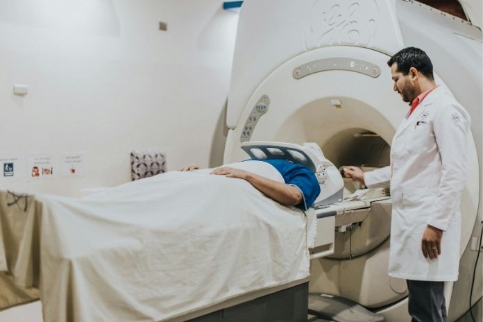
What to anticipate during the test is as follows:
- You will be required to lie on a bed that travels through a scanner that resembles a tunnel. Both ends of the scanner are open.
- During the scan, you must remain still, and the technician may ask you to hold your breath briefly.
- To take better photos, you might have a tiny gadget on your stomach or chest.
- During the process, a blood pressure cuff may be placed on your arm to monitor blood pressure.
- The scan can take up to ninety minutes, but if you need to talk to the person running the scanner (radiographer), you can hit a bell.
- The scanner makes a lot of noise. During the scan, you may hear loud tapping sounds. Usually, you can use earbuds or earplugs to listen to music.
Are Cardiac CT Scans and MRIs Safe?
When medically required, both cardiac CT scans and cardiac MRIs are usually safe. Although CT requires radiation exposure, the dose is greatly reduced by contemporary equipment. MRI does not use radiation, and doctors may use contrast dye carefully checked for kidney safety when needed.
Pacemakers and other implanted devices are important for the safety of noninvasive heart tests. Before prescribing imaging during pregnancy, clinicians carefully consider the hazards.
Limitations of Cardiac CT and MRI
Both heart imaging tests have limits despite their potency. They might need additional testing because they cannot predict every heart issue. Occasionally, incidental findings emerge that require more assessment. Even the results of an MRI or coronary CT angiography must be interpreted in conjunction with symptoms and other tests, such as an echocardiogram comparison.
How to Prepare for a Heart Imaging Appointment

Fasting, abstaining from caffeine, and dressing comfortably are some ways to prepare for a cardiac CT scan or cardiac MRI. Patients should inform their physicians about any contrast dye allergies and medications they are taking. It is also helpful to ask questions about the procedure, especially regarding what a cardiac CT scan shows or what a cardiac MRI is.
Read More: Bariatric Surgery Recovery: How to Hit Protein Goals on a Clear Liquid Diet
Conclusion
Two of the most useful instruments in contemporary cardiology are the cardiac CT scan and cardiac MRI, which provide extremely comprehensive pictures of the heart and blood vessels without requiring invasive procedures.
Cardiac imaging tests use both cardiac MRI and cardiac CT to help physicians identify problems with the arteries or heart muscle. Cardiac MRI provides the doctor with detailed information about the health of both the heart and any associated tissue.
Cardiac CT scans give a doctor the ability to quickly look for plaque buildup in the coronary arteries. Neither test is considered better than the other; rather, the choice is determined by the patient’s symptoms and clinical needs.
References
- South Tampa Cardiology. (2023, April 11). Cardiac MRI vs cardiac CT scans: Understanding the differences.
- Weill Cornell Medicine. Cardiac CT and MRI.
- WakeMed Health & Hospitals. Cardiac CT and MRI.
- University of Chicago Medicine. Cardiac CT angiography.
- Westchester Medical Center Health Network. Cardiac CT scan.
- Northwestern Medicine. (January 2024). What is cardiac MRI?
- British Heart Foundation. (25 September 2023). Cardiac MRI scan.
- Nebraska Medicine. What’s the difference between cardiac MRI and cardiac CT.
- Life Imaging Fla. Checklist before a heart scan.
In this Article