





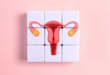













Most people can point to the clitoris on a diagram and stop there, satisfied that they understand it. That confidence is usually misplaced. For generations, anatomy textbooks reduced one of the body’s most nerve-dense organs to a tiny dot. Understanding clitoris anatomy means looking beyond the visible tip and recognizing a much larger internal structure.
The visible portion, called the glans, is only one small part of the clitoris. Most of the organ lies beneath the vulvar surface, forming a network of erectile tissue, nerves, and internal branches. Modern imaging studies have helped reveal how extensive this anatomy actually is. That research has challenged decades of incomplete medical understanding.
This lack of awareness has shaped everything from sex education to healthcare conversations. Many people still grow up learning very little about how the clitoris functions or why it matters. Misunderstandings about its anatomy can also affect conversations around pleasure, pain, arousal, and sexual health conditions. Even today, some myths continue because the organ was historically under-researched.
This article explains what the clitoris actually is, maps its complete anatomy, and explores how it responds during arousal. It also breaks down common myths and looks at conditions that can affect sensation, comfort, or appearance. Finally, it covers how hormones, aging, blood flow, and nerve health influence clitoral function over time. When necessary, it also explains when symptoms deserve medical attention.
- The clitoris is far larger than its visible tip, with most of its erectile tissue, including the body, crura, and bulbs, sitting internally beneath the vulva and around the vagina.
- A 2023 histomorphometric study counted roughly 10,280 nerve fibers in the dorsal nerve supplying the glans, about 20 percent above the long-quoted figure of 8,000.
- Modern imaging and cadaveric dissection corrected centuries of textbook neglect, reshaping how surgeons protect pelvic nerves and how clinicians counsel patients.
- Hormonal shifts, chronic pain, childbirth, pelvic surgery, and certain medications can all alter clitoral sensation, and persistent changes deserve medical evaluation.
What Is the Clitoris?
The clitoris is the primary organ of female sexual pleasure, and unlike the reproductive structures around it, that is essentially its whole job. It sits within the vulva, the external genitalia, and connects to a network of erectile tissue and nerves that extends well below the surface. Treating it as a single button misrepresents both its size and its function.
The part most people recognize is the glans, a small, rounded tip near the top of the vulva. That visible portion is only a fraction of the organ. Beneath the skin, the clitoris continues as a body and shaft, then splits into paired legs and bulbs that wrap toward the vaginal walls.
Research has put hard numbers on this hidden scale. The full structure measures roughly 9 to 11 centimeters when its internal components are included, a span no simple external diagram captures.
If you are wondering where the clitoris is located, the glans sits at the top of the vulva, above the urethral opening and the vaginal entrance, usually partly covered by a fold of skin called the clitoral hood. Its internal arms then arc backward and downward along the pubic bones on either side of the urethra and vagina.
Anatomy varies from person to person. The size of the glans, the extent of the hood, and the exact positioning of the internal structures differ, which is part of why sensation and preferred stimulation are so individual. None of that variation is abnormal.
Functionally, the clitoris exists for sexual pleasure. It is densely supplied with nerves and built from erectile tissue that engorges with blood during arousal, much like the penis to which it is embryologically related. Both develop from the same fetal tissue.
This sets it apart from organs with reproductive roles. The vagina serves childbirth and menstruation, and the uterus carries pregnancy, but the clitoris has no reproductive function. It is the body’s only structure dedicated solely to pleasure.
The Complete Anatomy of the Clitoris
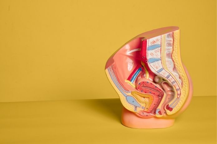
Mapping clitoris structure and function requires moving from the surface inward. Anatomists generally describe several connected parts: the glans, the hood, the body and shaft, the paired crura, and the vestibular bulbs. Each contributes to sensation, arousal, or both.
The Glans: The Visible External Portion
The glans is the only part of the clitoris outside the body. It is small, often a few millimeters to a centimeter in size, and exquisitely sensitive because of its dense innervation. For many people, it is the focal point of clitoral stimulation anatomy, though direct pressure can feel uncomfortable rather than pleasurable.
The clitoral hood, formed where the labia minora meet, drapes over the glans. It protects the tip from constant friction and shields the concentration of clitoral nerve endings underneath. How much of the glans the hood covers varies widely between individuals.
The Internal Body and Shaft
Just behind the glans, the clitoris continues as a short shaft and then a body, both made of erectile tissue. These run inward and upward before the structure divides. Most of this is impossible to see and easy to overlook, which is exactly why generations of diagrams left it out.
The Crura, or Legs, of the Clitoris
The body splits into two crura, often described as legs, that extend internally like a wishbone. Each leg runs several centimeters along the underside of the pubic bone, flanking the urethra and the vaginal canal. They are composed of corpora cavernosa, the same spongy erectile tissue found in the shaft.
The Vestibular Bulbs
Alongside the crura sit the vestibular bulbs, two masses of erectile tissue that flank the vaginal opening. During arousal, they fill with blood and swell, which puts gentle pressure on surrounding structures and contributes to the sensations of climax.
The bulbs sit in intimate contact with the urethra and the lateral walls of the vagina. This shared territory is why stimulation often does not feel cleanly separated by region, a point that matters for understanding the clitoris vs vagina debate later on.
Why Modern Scans Changed Scientific Understanding
For most of medical history, the internal clitoris anatomy was either ignored or actively minimized in teaching texts. Detailed neuroanatomy existed for the penis, while the clitoris went undescribed, a gap one pioneering researcher set out to correct.
Australian urologist Helen O’Connell, the first woman to train as a urologist in Australia and New Zealand, used MRI and cadaveric dissection to publish a full anatomical account in 2005. She has said her textbooks angered her because they described the penis in detail yet hardly mentioned clitoral anatomy at all. Her imaging work on clitoral anatomy reshaped both clinical and lay understanding of the organ.
Read More: Endometriosis Symptoms: Signs It Might Be More Than Just Painful Periods
How the Clitoris Works During Arousal
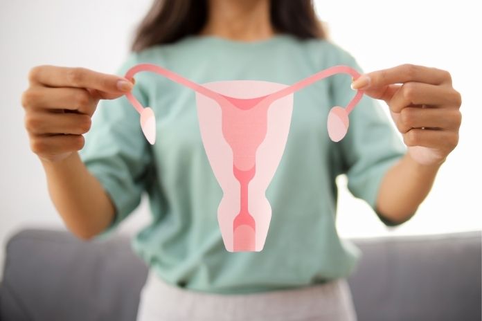
Arousal is a coordinated physical process, and the clitoris is at its center. Blood flow, nerve signaling, and the surrounding pelvic tissues all participate, which is why the response feels so different from one person to the next.
Blood Flow and Erectile Tissue Response
When a person becomes aroused, parasympathetic nerve signals increase blood flow to the erectile tissues of the penis. The corpora cavernosa of the shaft and crura, along with the vestibular bulbs, engorge and stiffen. The glans becomes more prominent, and the whole structure grows firmer, a female parallel to penile erection.
The Role of Nerve Endings
The clitoris is sensitive because of an extraordinary density of nerves. A 2023 study using actual tissue counting found that the dorsal nerve of the glans contains about 10,280 fibers, roughly 20 percent more than the 8,000 figure that has been repeated for years without a reliable source.
Maria Uloko, a urologist and assistant professor at the University of California, San Diego, who co-authored that study, has noted that such knowledge gaps in vulvar health produce real consequences for patients. Accurate numbers matter for surgery, nerve repair, and counseling.
How Stimulation Differs Between Individuals
Because anatomy and innervation vary, so does what feels good. Some people respond to light, indirect touch through the hood, others to firmer or rhythmic pressure. Speed, location, and intensity all shift the experience, and preferences can change with mood, hormones, and arousal level.
There is no single correct response pattern. The idea that everyone should react the same way to the same touch is a myth, not a clinical standard, and it sets up false expectations.
The Connection Between the Clitoris, Vagina, and Pelvic Floor
The clitoris does not operate in isolation. Its internal arms and bulbs sit against the vaginal walls and urethra, and the pelvic floor muscles contract during orgasm. Shared blood supply and nerve pathways link these structures into one functional system.
Blair Peters, a surgeon and assistant professor at Oregon Health & Science University who led the nerve-counting study, has pointed out that the widely cited 8,000 figure was never even an actual scientific paper. The point underscores how much of what people believe about this anatomy rests on repetition rather than evidence.
Common Myths About the Clitoris
Few organs attract as much misinformation. Some myths come from outdated textbooks, others from cultural discomfort with talking about female pleasure plainly. Here are the ones worth correcting.
This is the most persistent error. The visible glans is small, but the organ as a whole, counting the internal body, crura, and bulbs, is several inches in total span. Calling the clitoris tiny describes only the part you can see.
Researchers increasingly view this distinction as artificial. Because the internal clitoris surrounds the vaginal canal, stimulation described as vaginal often involves the clitoral structures indirectly. A 2013 anatomical review in Clinical Anatomy argued that the erectile tissues responsible for orgasm are essentially a single connected system rather than two unrelated mechanisms.
Anatomy and sensitivity differ enough that uniform expectations make little sense. What reliably produces pleasure for one person may do nothing for another, and that is a feature of normal human variation, not a defect.
Not reaching orgasm reliably is common and often has nothing to do with disease. Stress, fatigue, relationship dynamics, hormones, and medications all play roles. Treating every instance as a medical failure adds pressure that can make things worse.
Read More: How Stress Affects Your Menstrual Cycle — and What You Can Do About It
Why So Many People Were Never Taught the Full Anatomy
The knowledge gap is historical, not accidental. Female sexual anatomy was understudied, underfunded, and frequently treated as taboo, and the consequences reach into clinics today.
Medical training long centered male bodies as the default. Detailed accounts of penile nerves and blood supply existed while the clitoris was left largely undescribed, which is precisely the imbalance that motivated later researchers to start measuring and mapping.
Many classic texts drew the clitoris as a small external nub or omitted its internal structure entirely. Students absorbed an incomplete picture, then carried it into practice. Correcting that record required new imaging and a willingness to revisit assumptions.
When formal education skips accurate anatomy, people fill the gap with myth and guesswork. That leaves many adults unsure about basic female sexual anatomy and reliant on unreliable sources for information that affects their health and relationships.
Accurate anatomy is not trivia. It guides how surgeons operate in the pelvis to avoid damaging the pudendal nerve and its branches, informs the assessment of sexual pain, supports pelvic health care, and improves how clinicians and patients communicate about symptoms.
Medical Conditions That Can Affect the Clitoris

A range of conditions can change clitoral sensation, comfort, or appearance. Most are manageable, but they are worth recognizing rather than ignoring.
Declining estrogen during and after menopause thins genital tissues and reduces blood flow, which can lower sensitivity and contribute to discomfort. These changes are part of the broader genitourinary syndrome of menopause and are treatable.
Some people develop persistent clitoral or vulvar pain, sometimes involving the pudendal nerve, and others experience heightened, unwanted sensitivity. These conditions are real, can be distressing, and deserve evaluation by a clinician familiar with sexual medicine.
Vaginal delivery, episiotomy, and surgical procedures in the pelvic region can affect nearby nerves and tissues. This is one reason detailed anatomical knowledge matters: protecting the clitoral nerves during surgery depends on knowing where they actually run.
Certain drugs blunt sexual response. Selective serotonin reuptake inhibitors and other antidepressants are well known for reducing arousal and delaying or preventing orgasm. Hormonal medications can also shift sensitivity. Anyone troubled by these effects can discuss alternatives with a prescriber rather than simply enduring them.
Read More: More Than “In Your Head”: 5 Physical Signs Relationship Anxiety Is Affecting Your Body
When to Talk to a Healthcare Professional
Changes in sexual sensation are common, but some warrant attention. Knowing when to raise a concern helps people get answers instead of quietly worrying.
New pain, numbness, or a clear shift in how the clitoris responds is worth mentioning to a clinician, ideally a gynecologist or someone versed in sexual medicine. Such changes can have treatable causes, from infection to nerve issues to hormonal shifts.
Ongoing discomfort during sex, or distress about sexual function, is a legitimate medical concern. It is not something to dismiss as purely psychological or unimportant.
Anyone noticing altered sensation after a pelvic procedure, an injury, or delivery should flag it. Early conversation can identify whether the change is expected, temporary, or treatable.
Sexual health is part of overall health. Rachel Rubin, a urologist and sexual medicine specialist, has said that gaps in clinicians’ basic anatomical knowledge can lead to harm and intense sexual dysfunction that she sees in her practice. Reducing stigma and asking questions improves quality of life.
How Better Understanding of the Clitoris Supports Overall Sexual Health
Knowledge changes how people relate to their own bodies and how they advocate for care. Accurate information about clitoris anatomy is a foundation for both.
Understanding the full structure makes it easier to describe what feels good and what does not, for both partners and clinicians. That awareness reduces frustration and supports healthier intimacy.
When people know how arousal and sensation actually work, they hold more realistic expectations and waste less energy worrying about being abnormal. Accurate anatomy is quietly reassuring.
Online spaces are full of confident claims that contradict the evidence. Grounding understanding in research and clinical sources protects people from misinformation that can shape decisions about their health.
Key Takeaway: The Clitoris Is a Complex Organ, Not Just a Small External Structure
The clitoris is a large, intricate organ with external and internal components, built from erectile tissue and supplied by thousands of nerve fibers. Reducing it to a visible button, as so many diagrams have, misses nearly all of what it is. Understanding clitoris anatomy starts with accepting that most of it lies beneath the surface.
Scientific understanding has improved dramatically, from O’Connell’s imaging work to the first real count of clitoral nerve fibers, yet education has not fully caught up. Many people still learn an outdated version, and that gap has consequences for how they understand their bodies and seek care.
The practical response is straightforward: approach sexual health with curiosity, rely on accurate information rather than myth, and treat concerns about sensation or function as worth a conversation with a qualified clinician. Better knowledge of the clitoris supports real health and wellbeing.
References
- O’Connell, H. E., Sanjeevan, K. V., & Hutson, J. M. (2005). Anatomy of the clitoris. The Journal of Urology, 174(4 Pt 1), 1189–1195.
- Pauls, R. N. (2015). Anatomy of the clitoris and the female sexual response. Clinical Anatomy, 28(3), 376–384.
- Puppo, V. (2013). Anatomy and physiology of the clitoris, vestibular bulbs, and labia minora with a review of the female orgasm and the prevention of female sexual dysfunction. Clinical Anatomy, 26(1), 134–152.
- Uloko, M., Isabey, E. P., & Peters, B. R. (2023). How many nerve fibers innervate the human glans clitoris: A histomorphometric evaluation of the dorsal nerve of the clitoris. The Journal of Sexual Medicine, 20(3), 247–252.
- Oregon Health & Science University. (2022, October 27). Pleasure-producing human clitoris has more than 10,000 nerve fibers. OHSU News.
- PBS NewsHour. (n.d.). Why is women’s sexual health so understudied?
- Cleveland Clinic. (n.d.). Clitoris. Cleveland Clinic.
- O’Connell, H. E., Sanjeevan, K. V., & Hutson, J. M. (2005). Anatomy of the clitoris. The Journal of Urology, 174(4 Pt 1), 1189–1195.
- Clue. (n.d.). What is the clitoris? Clue.
- Kenhub. (n.d.). Clitoris: Anatomy and function. Kenhub.
- Wikipedia contributors. (n.d.). Clitoris. In Wikipedia. Retrieved May 29, 2026
- Edwards, B. A. (2023, November 13). “Anatomy of the clitoris” (2005) by Helen E. O’Connell, Kalavampara V. Sanjeevan, and John M. Hutson. The Embryo Project Encyclopedia.
- Metropolis Healthcare. (n.d.). Clitoris anatomy and function. Metropolis Healthcare.
In this Article