

















Not all breast lumps are cancerous; some are benign and disappear on their own. Breast lumps are mainly fibroadenomas, cysts, and fibrocystic changes. However, in some cases, breast lumps can be cancerous too. Benign breast lumps feel smooth, mobile, and consistent with the menstrual cycle. Malignant lumps are hard, fixed, painless, and have irregular borders. But you cannot determine whether a breast lump is benign or malignant by physical examination or imaging. It needs further diagnosis.
In this article, we discuss the major differences between a benign breast lump and cancer, their characteristics, red flags you should be aware of, and why imaging plays a key role in identifying the type of breast lump and cancer.
- A breast lump is a mass or growth found in your breast.
- Most of the breast lumps are benign but might look like cancer.
- If you find one on your breast or near your armpit, it is recommended to see a healthcare provider.
Read More: Breast pain before periods
Types Of Benign Breast Lumps Similar to Cancer
The following are the types of breast lumps commonly found in women.
Fibroadenoma
Fibroadenoma is common in women above 30, which occurs during puberty, the menstrual cycle, or through hormonal fluctuations. They are typically present in the upper outer quadrant of the breast and feel smooth, firm, and mobile.
These lumps are composed of glandular and fibrous tissue and do not cause significant breast pain or fluctuate with the menstrual cycle.
Breast Cysts
There are 3 types of cysts.
- Simple cysts: These cysts appear as fluid-filled sacs with smooth, thin, regularly shaped walls.
- Complex cysts: Have irregular or scalloped borders and thick walls and contain fluid as well as some solid areas.
- Complicated cysts: These cysts may appear solid on imaging and require careful evaluation to differentiate them from complex cysts. In complex cysts, there is a 20 to 30% chance of malignancy.
Phyllodes Tumour
Phyllodes tumors can be benign or malignant, and there are a few cases in which they fall into a borderline category.
It is difficult to differentiate between fibroadenomas and Phyllodes tumors even on mammogram or imaging, as they start off as painless masses in the connective tissue in the breast.
Fibroadenomas usually resolve on their own and do not require treatment. But Phyllodes Tumor
should be removed as they tend to grow quickly, even when not cancerous.
Fat Necrosis
Fat Necrosis is usually benign but can act as breast cancer. Thus, clinicians and radiologists recommend imaging modalities such as mammography, ultrasound, and MRI.
The skin around the lump may look thicker, redder, or bruised, similar to what is seen with breast cancer.
If a woman has fat necrosis, she might also notice skin tethering, dimpling of the overlying skin, and nipple retraction, which again resembles malignancy.
Mastitis and Breast Abscess
When a woman is affected with Mastitis and a breast abscess, it can cause redness, warmth, and skin thickening, tenderness, and a palpable lump.
A doctor’s intervention is necessary if you experience any of the symptoms listed above. Matitis might act similarly to inflammatory breast cancer. If symptoms do not improve with medication, an investigation for cancer is necessary.
Note: Fibroadenomas and some cancers feel identical on examination. Therefore, all new discrete breast lumps must undergo imaging regardless of their clinical features.
Read More: Breast health essentials
Breast Cancer Lump vs Benign Features
Here’s how to differentiate a breast cancer lump from a benign breast lump through its features. Remember, despite these features, a breast lump can be confirmed as benign or malignant through biopsy.
Health & Screening
Benign Breast Lump vs Cancer — Physical Features
| Feature | Benign | Cancerous | Caveat |
|---|---|---|---|
| Texture Hard vs soft |
Soft / rubbery | Hard / stony | Some cancers feel soft. Some benign lumps feel very firm. |
| Mobility Moves vs fixed |
Moves freely | Fixed to tissue | Early-stage cancers can still feel mobile. Fixed = advanced cancer only. |
| Borders Smooth vs irregular |
Smooth, defined | Irregular, spiky | Borders are best assessed on ultrasound — not reliably felt on examination. |
| Pain Tender vs painless |
Often tender | Usually painless | ~10% of breast cancers do cause pain. Painless ≠ safe. |
| Size change Growth rate |
Stable / slow | Rapid growth | Size alone is not diagnostic. Rapid growth in any lump warrants reassessment. |
Read More: 10 Breast Cancer Myths
Signs Breast Lump Could Be Cancerous
Breast cancer is only diagnosed in about 10% of new breast lump presentations. But you must seek immediate medical attention if you notice the following symptoms:
Skin Changes
If the overlying skin appears dimpled, puckered, or has an orange-peel texture (peau d’orange). These signs could suggest malignancy.
Fat necrosis can occasionally lead to skin dimpling. That is why performing a biopsy remains essential to differentiate one type of lump from another.
Nipple Changes
If the nipple is turning inward, becoming retracted, or changing position, it can sometimes be a warning sign of an underlying breast tumor.
Nipple Discharge
Bloody nipple discharge is most commonly caused by intraductal papilloma and duct ectasia when compared to breast cancer. However, nipple discharge is also associated with a 5-23% risk of malignancy.
Discharge due to malignant disease is significantly more likely to be blood-stained than discharge due to benign causes.
If you notice discharge is spontaneous and not expressed, unilateral, from a single duct, blood-stained or clear, then it needs further investigation.
Axillary Lymph Node Swelling
If you observe firm and non-tender lumps in the armpits in addition to a breast lump, this could mean cancer has spread to the regional lymph nodes. In some cases, seeking professional help is a must.
Inflammatory signs
Inflammatory breast cancer is the most aggressive form of cancer, and its detection is hard. Inflammatory breast cancer cells block lymph channels in the skin rather than forming a distinct lump, making it one of the hardest breast cancers to detect early.
When someone is infected with inflammatory breast cancer, they might notice redness, warmth, swelling of the entire breast, or rash covering more than 1/3rd of the breast.
Any Lump in a Postmenopausal Woman
The risk of a breast lump being malignant rises with age. A woman in her thirties has roughly a 1 in 204 (0.49%) chance of a lump being cancer, compared to a 1 in 28 (3.54%) chance for a woman in her sixties. If there’s a lump after menopause, all new breast lumps demand further investigation and imaging.
Read More: What early-onset breast cancer feels like
How Imaging Distinguishes Benign From Malignant?
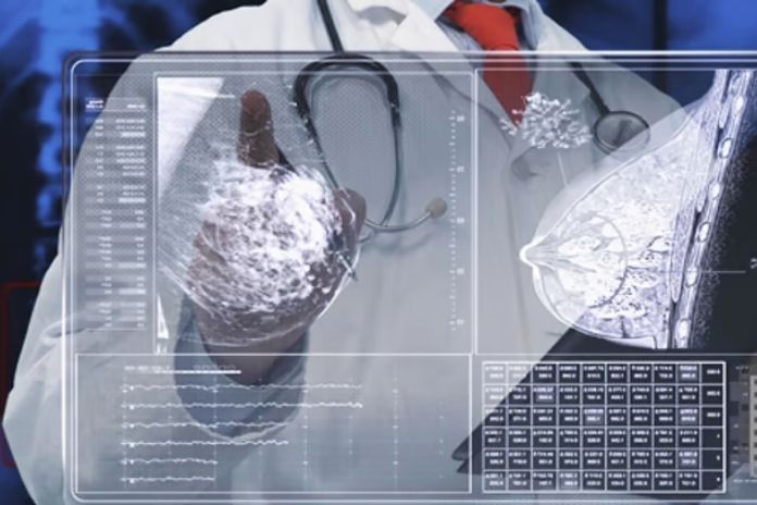
While physical examination establishes a clinical impression, imaging provides a probability, and biopsy yields a diagnosis.
Mammography
Mammography is a primary screening performed on women over 40 to detect cancer or other breast lump types, like fibroadenomas or cysts.
Ultrasound
Ultrasound is performed as initial imaging for women under 35 to evaluate palpable lumps. Ultrasound allows doctors to differentiate simple cysts from solid masses and to assess borders, texture, and other features.
If the breast lump is known to be benign, it appears to have a smooth, oval outline. If there are irregular margins and an increased blood supply, then it could be cancer.
BI-RADS Classification
Radiologists use the standardized Breast Imaging Reporting and Data System (BI-RADS) scale, ranging from 0 to 6, to communicate the probability of malignancy. BI-RADS 3 means probably benign (a six-month follow-up is recommended). BI-RADS 4 means suspicious (biopsy recommended). BI-RADS 5 means highly suggestive of malignancy.
Core Biopsy
A biopsy is a definitive test to confirm malignancy. It identifies cancer by removing a sample of tissue, cells, or fluid, which a pathologist then examines under a microscope.
Depending on the tumor location, the procedure varies. It could include needle aspiration, endoscopic, skin, or surgical removal.
When to See a Doctor?

You must consult a doctor when you find a new, discrete lump. A growing lump. A lump in a postmenopausal woman. And if the lump is associated with skin changes, nipple inversion, discharge, or axillary swelling.
If a lump was previously found to be benign, it is necessary to follow up and continue regular self-examinations and routine screening.
If you notice redness, swelling, and warmth around your breasts without any signs of infection, go to the emergency ward. This can indicate inflammatory breast cancer, which acts similarly to an infection but does not subside with antibiotics.
NOTE: Do not delay examination of a lump for over a week after noticing it. This is because any lump can be confirmed with imaging.
Read More: What healthy practices should breast cancer patients follow
Conclusion
When you notice a breast lump, do not be frightened; most of them are benign, and a few of them disappear on their own. That said, if the lump persists for more than a week, it needs a physical examination.
If you find redness, warmth, a lump spreading to surrounding areas, or blood stains on your nipple, or a change in nipple position, seek immediate care.
Finally, ensure you follow the procedure thoroughly, as your doctor suggests, whether the condition is benign or malignant. Treating breast lumps on time can be successful.
FAQs
How do you know if a breast lump is cancerous without a biopsy?
Without a biopsy, there is no definitive way to confirm cancer. But you can determine the probability through BI-RADS. A biopsy is necessary, as breast lump types are similar or overlap with their characteristics, making it difficult to analyze through examination or imaging.
Are cancerous breast lumps always hard?
No. Although hard lumps mostly indicate malignancy rather than benignity. But there are other factors, like examining the surrounding tissue rather than the texture alone. In this case, the lump is benign; it is firm, whereas some cancers are soft to the touch. Other factors to consider for physical examination include texture, mobility, and surrounding tissue.
Can a breast lump be cancerous if it moves?
No. Both early-stage cancers and fibroadenomas can feel mobile, although fibroadenomas are benign. Professionals can confirm cancer through imaging and biopsy.
What is the most important change that suggests a breast lump might be cancer?
Changes that indicate cancer include skin dimpling or puckering over the lump; nipple inversion or retraction; a fixed, rather than mobile, consistency; and axillary lymph node swelling alongside the lump. Any one of these warrants urgent assessment.
Blood-stained spontaneous nipple discharge is also highly significant. Of all the changes that can occur, skin changes are the most specific clinical indicator that a deeper structural abnormality, likely malignant, is affecting the overlying tissue.
In this Article